Last updated: 23 October 2026
If you are having follicle-tracking scans, your report will often mention a dominant follicle and a size in millimetres. Those numbers can be confusing, especially when you are trying to conceive and every scan feels important. This guide explains what it is, the usual follicle size for ovulation, why the day of the cycle matters, and what to discuss with your doctor.
Remember. Follicle size is a guide to timing, not a test of egg quality or a prediction of pregnancy. Your doctor reads it together with your cycle, hormone results and the lining of the womb.
What is a follicle, and what does dominant follicle mean?
A follicle is a small fluid-filled sac in the ovary that contains an immature egg. Several small (antral) follicles start to grow early in each cycle. Usually one grows faster than the others and the rest stop growing. That leading follicle is the dominant follicle, and in a natural cycle it is usually the one that releases the egg at ovulation.
So the dominant follicle meaning, in simple terms, is “the follicle most likely to ovulate this cycle”. StatPearls describes a follicle as dominant once it is larger than about 10 mm, usually by around day 8 to 12 of the cycle.
How a follicular scan (follicle tracking) is done
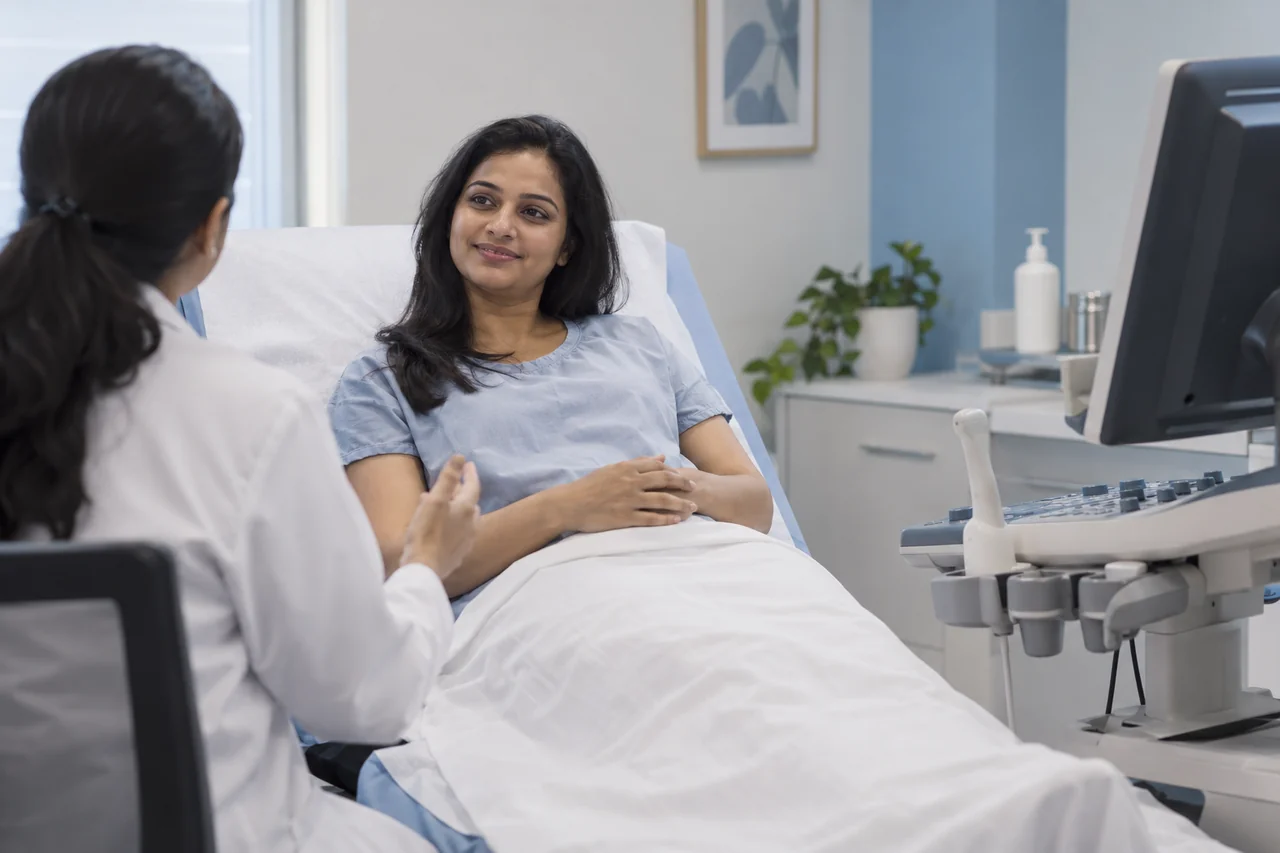
Follicle tracking, also called folliculometry, is a series of transvaginal ultrasound scans. Patient information from ISUOG describes it as a simple outpatient scan that takes about 10–15 minutes. For a natural or IUI cycle, scans often start around day 9–10; the exact days depend on your cycle length and treatment plan. A first scan early in the cycle may also be done to check the ovaries and lining. Follicle-tracking scans are done at our centre in Kompally, as part of ovulation induction and IUI treatment in Kompally.
Follicle size for ovulation: the usual range
Published patient information gives slightly different ranges, which is normal because people and cycles vary:
| Source | What it says about follicle size |
|---|---|
| ISUOG (folliculometry) | Ovulation usually happens when the leading follicle is about 18–22 mm; follicles grow about 1–2 mm per day |
| ASRM ReproductiveFacts | Ovulation generally happens when the follicle measures about 1.8–2.5 cm (18–25 mm) |
| StatPearls (NCBI) | In a natural cycle, the leading follicle grows about 1–1.4 mm per day to a mean of about 20–24 mm at ovulation; other studies report a wider range |
| NHS patient leaflet (Ashford and St Peter’s) | By about day 12–14, the follicle should have reached roughly 17–25 mm |
In other words, the egg follicle size at ovulation is usually somewhere around the high teens to mid-20s in millimetres. A single measurement slightly outside these numbers is not, by itself, a problem. Your doctor looks at how the follicle grows over several scans.
Normal follicle size on day 14
People often search for the normal follicle size on day 14. Day 14 is only “mid-cycle” in a 28-day cycle. If your cycles are longer, ovulation usually comes later, so a smaller follicle on day 14 can be perfectly normal for you. That is why follicle tracking is about the trend over several days, not one day’s number.
Follicle size in treatment cycles
When tablets or injections are used to help ovulation, more than one follicle may grow and the growth rate can differ. A trigger injection is often used to time ovulation. Yale Medicine describes giving the trigger once the lead follicle is at least about 16–18 mm, and StatPearls notes that many IVF centres use about 18–20 mm before egg collection. Protocols vary, so your doctor decides the timing for your cycle. If several large follicles grow, your doctor will also talk to you about the risk of a multiple pregnancy before going ahead.
Minimum egg size to get pregnant: a common mix-up
The phrase “minimum egg size to get pregnant” usually refers to follicle size, not the egg itself. The egg is microscopic and cannot be measured on an ultrasound scan. What the scan measures is the fluid-filled follicle around it. There is no single cut-off that decides whether pregnancy is possible; the size ranges above are used to time ovulation, IUI or egg collection.
What does “no dominant follicle” mean?
If a scan shows none, it means that on that day no follicle had grown ahead of the others. It may simply be too early in the cycle, or that cycle may be one where ovulation does not happen (an anovulatory cycle). Possible reasons your doctor may look into include PCOS, thyroid or prolactin problems, low ovarian reserve (see understanding AMH), recent weight change or stress, and some medicines. One such cycle does not mean you cannot ovulate; your doctor may repeat the scan, check hormones or adjust treatment.
Leading follicle in the right ovary: does it mean pregnant?
No. Which side the leading follicle is on does not show whether you are pregnant, and it does not change your chance of pregnancy in any reliable way. Either ovary can ovulate in a given cycle. Pregnancy can only be confirmed by a pregnancy test after the expected period, and later by a scan.
What else your scan report may mention
- Number and size of follicles in each ovary — usually listed in millimetres.
- Endometrium (lining of the womb) — its thickness and appearance, which your doctor reads alongside follicle growth.
- Signs of ovulation — on a later scan, the follicle may have collapsed and formed the corpus luteum.
- Cysts or other findings — these are explained by your doctor, as they may change the plan.
What a follicle scan cannot tell you
- The quality of the egg inside the follicle.
- Whether the fallopian tubes are open — that needs a tubal test such as HSG.
- The male partner’s fertility — that needs a semen analysis.
- An exact chance of pregnancy in that cycle.
Questions to ask at your follicle-tracking visit
- How many follicles are growing, and how big is the leading one?
- Is the growth on track for my cycle length?
- When is the next scan, and will a trigger injection be needed?
- When should we plan intercourse or IUI?
- Will you check later whether ovulation happened?
Follicle monitoring at Sree Nandaka IVF, Kompally
At Sree Nandaka IVF Centre, follicle-tracking scans are planned around your cycle and treatment, and the results are explained to you by Dr. Deepa Rekha Koduru, Fertility Specialist (MBBS, DGO; DRM, Diploma in Reproductive Medicine, Germany; MRM, Master in Reproductive Medicine, Homerton University, London; Reg. No. 54076). If IUI is part of the plan, our guide to what happens after IUI explains the next two weeks. For a wider check-up, see our fertility evaluation page.
Frequently asked questions
It is the one that grows ahead of the others in a cycle. In a natural cycle it is usually the one that releases the egg at ovulation.
Patient information from ISUOG gives about 18–22 mm, and ASRM gives about 18–25 mm. Cycles vary, so your doctor looks at growth over several scans rather than one number.
It depends on your cycle length. In a 28-day cycle the leading follicle is often in the high teens to low 20s in millimetres around day 12–14, but with longer cycles ovulation usually comes later.
On that day no follicle had grown ahead of the others. It may be too early in the cycle, or ovulation may not happen in that cycle. Your doctor may repeat the scan or check hormones.
No. The side of the leading follicle does not show pregnancy. Pregnancy is confirmed by a pregnancy test after the expected period, and later by a scan.
The egg itself cannot be measured on a scan; the size on the report is the follicle. Follicle size is used to time ovulation, IUI or egg collection, not to decide whether pregnancy is possible.
Conclusion
The leading follicle is the one most likely to release an egg in that cycle. The usual follicle size for ovulation is roughly the high teens to mid-20s in millimetres, but the trend across several scans matters more than one number, and your cycle length changes what is “normal” on a given day. Ask your doctor to explain your scan together with your hormones, lining and partner’s report.
Book a consultation at our Kompally centre
Sree Nandaka IVF Centre, Plot 770, Jayabheri Park, behind Big Bazaar, Kompally, Hyderabad, Telangana 500014.
- Call +91 76720 01080
- Message us on WhatsApp +91 76720 01080 or email sreenandakafertility@gmail.com
- Book a Consultation online (MocDoc)
Please note. This article is general information, not medical advice. Scan findings and treatment timing depend on your history and a specialist assessment. No test or treatment can guarantee a pregnancy.
Sources
- ISUOG patient information: Folliculometry
- ASRM ReproductiveFacts: Am I Ovulating?
- StatPearls (NCBI Bookshelf): Sonography Gynecology Infertility Assessment, Protocols, and Interpretation
- Ashford and St Peter’s Hospitals NHS Foundation Trust: Follicle tracking scans (patient leaflet)
- Yale Medicine: Ovulation induction and intrauterine insemination
Written by the Sree Nandaka IVF Centre editorial team, Kompally. General information only — not a substitute for a consultation.